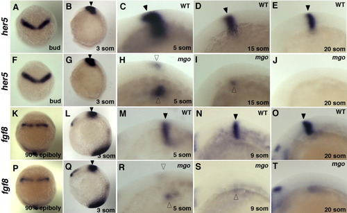
her5 and fgf8 expression profile in mgo mutants. Dorsal views (A, F, K, P) of embryos with anterior at the top; lateral views (B-E, G-J, L-O, and Q-T) with anterior to the left. A-J: Analysis of her5 expression in wild-type (C-E) and mgo mutants (H-J). At the bud and 3-somite stage, no difference was detected in her5 expression (A,B,F,G). By 5-somite stage, lower her5 expression was noted in mgo mutants (H). her5 expression remained only in the ventral region at the 15-somite stage and disappeared completely by the 20-somite stage (I,J). K-T: fgf8 expression in Wild-type (M-O) and mgo homozygous mutants (R-T). Expression of fgf8 initiated in all embryos within the MHB primordia at 90% epiboly and remained at the 3-somite stage (K, L, P, Q). By the 5-somite stage, fgf8 expression was reduced especially in dorsal region (R). Expression of fgf8 remained within the ventral region at the 9-somite stage (S) and was absent by the 20-somite stage (T). Developmental stages and genotype are indicated in the bottom and top right-hand corner, respectively. Open arrowheads point to mgo MHBs.
|
