|
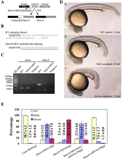
Targeted knockdown of Mtmr8 using splicing morpholino in zebrafish embryos. (A) Diagram of splicing junction morpholino targeted against Mtmr8 exon-intron boundary. (B) Amino acid sequences of wild type Mtmr8 and Mtmr8-MO1 mediated mis-splicing Mtmr8. Sequencing of the RT-PCR products revealed the mis-splicing transcript leading to a premature stop (asterisk) and causing a truncation in the protein (exon 1 in bold, the other exons in plain, and intron in plain and underlined). (C) RT-PCR detection of Mtmr8 transcript at 24hpf in WT and Mtmr8-MO1 (6 ng) morpholino-injected embryos, comparing cryptic spliced transcript in the morpholino injected embryos to the cDNA PCR products. (D) Live morphology of WT control zebrafish embryo (a) and Mtmr8-MO1 knockdown embryo (b, c) at 24hpf. Injection volume was about 2 nL at 1-cell stage embryos. All scale bars are 100 μm. (E) Statistical data of three independent experiments on Mtmr8 knockdown as well as its overexpression and Mtmr8 mRNA rescue. Results are represented as mean±SD of three separate experiments (60 embryos in each experiment).
|