
Fig. 9
- ID
- ZDB-FIG-090324-28
- Publication
- Kimmel et al., 1998 - The shaping of the pharnygeal cartilages during early development of the zebrafish
- Other Figures
- All Figure Page
- Back to All Figure Page
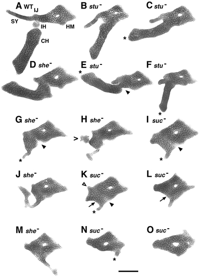
A phenotypic series in the shapes of mutant hyoid cartilages. Flat-mounted dissected preparations from stu, she, and suc mutants at day 5, arranged according to how much hyoid arch cartilage is present. A wild-type preparation is also included (A). The highly variable she- phenotype overlaps both stu- and suc-. Two she- examples (G, H) fall in the middle of the series. Another (D) fits ahead of stu mutants, and two (J, M) fit after one or more suc- examples. Our interpretation of the identities of particular regions of cartilage is explained in detail in the text. In contrast to disruption of the ventrolateral hyoid (CH, IH, SY) cartilages, the dorsal HM region is readily identifiable and reasonably normal in shape and size in every mutant we examined, even though it is nearly invariably fused to other cartilages. The assignment is based on the following features, shared with the HM in wild types: The mutant HM is present as a diamond-shaped region of cartilage that articulates with the otic capsule at its dorsal–anterior apex. The HM is located just posterior to a cartilage we interpret to be its mandibular arch homologue, the palatoquadrate (e.g., as shown in Fig. 10). Muscles insert to the mutant HM that from their origins on the chondrocranium seem to be the correct ones (see Schilling and Kimmel, 1997). The mutant HM invariably has a central hole, corresponding to the wild-type hyomandibular foramen. Finally, the mutant HM is often associated with a bone primordium we interpret to be the opercular, at a posterior–ventral apex of the HM as usual (example in Fig. 10). In all three mutants, ectopic small cartilages of unknown identity are often present (H; .), emphasizing the need for region-specific molecular markers that are only beginning to become available (Schilling, 1997). Asterisks (C, E, F, G, I, K, N) indicate the position we take to be the tip of the mutant CH. The triangle (K) indicates a stub of cartilage corresponding in position to the SY. Arrowheads (E, G, I, K) indicate where we propose the joint between the HM and the CH would normally form. Arrows (K, L) show chondrocyte stacks curving ventrally from the IJ. Such curving cell stacks are also present in other mutants in this part of the series (e.g., J) but are never observed in wild types (e.g., Fig. 2). Scale bar: 100 μm. |
| Fish: | |
|---|---|
| Observed In: | |
| Stage: | Day 5 |
Reprinted from Developmental Biology, 203, Kimmel, C.B., Miller, C.T., Kruse, G., Ullmann, B., BreMiller, R.A., Larison, K.D., and Snyder, H.C., The shaping of the pharnygeal cartilages during early development of the zebrafish, 245-263, Copyright (1998) with permission from Elsevier. Full text @ Dev. Biol.