
Fig. 9
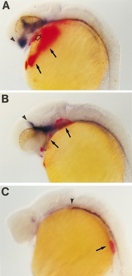
Cyclopic embryos lack correctly positioned hatching glands. Ethanol-treated embryos at the 24-h stage were simultaneously stained for hgg-1 (red) and shh (blue) expression; hgg1 is specifically expressed in hatching glands. (A) Ethanol-treated embryo with narrowly spaced eyes have hatching glands over the yolk (arrows). Some hgg1-expressing cells were incorrectly positioned under the forebrain (open arrow). (B) Cyclopic embryo which lacks the wild-type pattern of hgg-1 expression on the yolk; hatching glands are instead found at the level of the forebrain and midbrain (arrows). Note that the anterior-most expression of shh in the hypothalamus is missing in this embryo (compare arrowheads marking anterior limit of shh expression in A and B). (C) Profoundly cyclopic embryo which lacks shh expression in the fore- and hgg-1-expressing cells are located in the trunk in this embryo (arrow). Orientation of embryos is anterior to the left and dorsal up. Arrowheads, anterior limits of shh expression. |
Reprinted from Developmental Biology, 201, Blader, P. and Strähle, U., Ethanol impairs migration of the prechordal plate in the zebrafish embryo, 185-201, Copyright (1998) with permission from Elsevier. Full text @ Dev. Biol.