Fig. 6
- ID
- ZDB-FIG-090310-10
- Publication
- Buckles et al., 2004 - Combinatorial Wnt control of zebrafish midbrain-hindbrain boundary formation
- Other Figures
- All Figure Page
- Back to All Figure Page
|
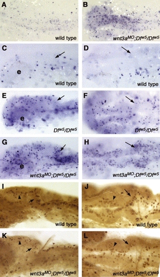
Wnt signaling is required to inhibit apoptosis in the brain. (A–H) TUNEL assays to detect apoptosis. (I–L) Assays of phospho-histone H3 labeling. All panels; anterior is to the left, genotype is indicated in lower right corner. (A,B) 18-somite stage embryos, dorsal view. Substantially increased numbers of apoptotic cells are observed. (C,E,G) Lateral views and (D,F,H) dorsal views of 24 hpf embryos. (C,D) Wild type embryos have very little cell death at this stage. e: eye. (E,F) Consistent with our previous report (Lekven et al., 2003), Dfw5/Dfw5 embryos have increased apoptosis in the tectum, but not in the cerebellum (arrows in C–L indicate cerebellum). (G,H) wnt3aMO;Dfw5/Dfw5 embryos have greatly elevated levels of apoptosis in the midbrain and cerebellum. (I,K) Lateral views and (J,L) dorsal views of 24 hpf embryos labeled for phospho-histone H3. (I,J) Wild type embryos have regular rows of mitotic cells at the dorsal midline of the midbrain (arrowheads) and in the rhombic lip (arrows). (K,L) wnt3aMO;Dfw5/Dfw5 embryos occasionally have reduced numbers of mitotic cells in these locations. |
| Fish: | |
|---|---|
| Knockdown Reagent: | |
| Observed In: | |
| Stage Range: | 14-19 somites to Prim-5 |
Reprinted from Mechanisms of Development, 121(5), Buckles, G.R., Thorpe, C.J., Ramel, M.C., and Lekven, A.C., Combinatorial Wnt control of zebrafish midbrain-hindbrain boundary formation, 437-447, Copyright (2004) with permission from Elsevier. Full text @ Mech. Dev.

