
FIGURE
Fig. 11
- ID
- ZDB-FIG-090304-86
- Publication
- Ma et al., 2009 - Establishment of a transitory dorsal-biased window of localized Ca(2+) signaling in the superficial epithelium following the mid-blastula transition in zebrafish embryos
- Other Figures
- All Figure Page
- Back to All Figure Page
Fig. 11
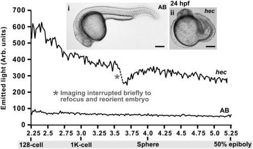
Representative changes in [Ca2+]i that occur in AB wild-type and hec mutant embryos during the Blastula Period (i.e., from the 128-cell stage to 50% epiboly). f-aequorin-generated data were plotted every 60 s, each data point representing 60 s of accumulated luminescence for an imaging field covering the entire blastoderm of each embryo (approximately 50,000 pixels). The traces are representative of n = 10 AB and n = 7 hec mutant embryos. The bright-field images (panels i and ii) show the morphology of the AB and hec embryos at 24 hpf. Scale bars are 200 μm. |
Expression Data
Expression Detail
Antibody Labeling
Phenotype Data
Phenotype Detail
Acknowledgments
This image is the copyrighted work of the attributed author or publisher, and
ZFIN has permission only to display this image to its users.
Additional permissions should be obtained from the applicable author or publisher of the image.
Reprinted from Developmental Biology, 327(1), Ma, L.H., Webb, S.E., Chan, C.M., Zhang, J., and Miller, A.L., Establishment of a transitory dorsal-biased window of localized Ca(2+) signaling in the superficial epithelium following the mid-blastula transition in zebrafish embryos, 143-157, Copyright (2009) with permission from Elsevier. Full text @ Dev. Biol.