
Fig. S2
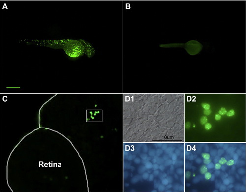
neuroDEGFP fusion protein is translated in vivo. Panel A illustrates a transient transgenic embryo treated with heat shock at 24hpf and photographed at 48hpf. Note the mosaic of GFP-labeled cells. Panel B is an uninjected control treated with heat shock. Panel C illustrates a section through the head and retina of a transient transgenic embryo at 48hpf. The enclosed area is illustrated at higher magnification (and rotated slightly) in D. Note the nuclear localization of the GFP. D1, Nomarski illumination; D2, GFP fluorescence; D3, nuclear staining with bisbenzimide; D4, digital overlay D2 and D3. The scale bar in A represents 200 μm for panels A and B and 50 μm for panel C. |
Reprinted from Mechanisms of Development, 126(3-4), Ochocinska, M.J., and Hitchcock, P.F., NeuroD regulates proliferation of photoreceptor progenitors in the retina of the zebrafish, 128-141, Copyright (2009) with permission from Elsevier. Full text @ Mech. Dev.