
Fig. 6
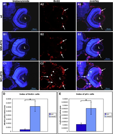
Cells fail to withdraw from the cell cycle following knock down of neuroD. Panels A, B and C illustrate retinas form wt, mismatch control and morphant fish, respectively, that were labeled with BrdU and sacrificed at 72hpf. Left-hand panels illustrate nuclear staining with bisbenzimide; middle panels illustrate BrdU labeling; right-hand panels are the respective digital overlays. L, lens. Panel D is histograms comparing the number of BrdU-labeled cells in control and morphant retinas. Panel E is histograms comparing the number of cells immunopositive for phosphohistone H3 in control and morphant retinas. Asterisks, p < 0.001. |
Reprinted from Mechanisms of Development, 126(3-4), Ochocinska, M.J., and Hitchcock, P.F., NeuroD regulates proliferation of photoreceptor progenitors in the retina of the zebrafish, 128-141, Copyright (2009) with permission from Elsevier. Full text @ Mech. Dev.