
Fig. 3
- ID
- ZDB-FIG-090304-37
- Publication
- Nevin et al., 2008 - Hardwiring of fine synaptic layers in the zebrafish visual pathway
- Other Figures
- All Figure Page
- Back to All Figure Page
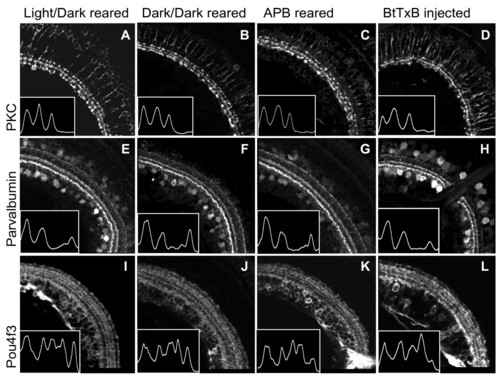
Dark-reared, APB-treated, and BtTxB-injected larvae show proper IPL sublamination.(A-L) Sections showing the IPL of 5 dpf larvae raised in a normal light:dark cycle (A, E, I), constant darkness (B, F, J), in the presence of 1 mM APB (C, G, K), and treated with BtTxB (D, H, L). The images in D, H, L are from the larva recorded in Figure 4D. Insets: traces of the fluorescent signal intensity across the width of the IPL (region shown). Peaks correspond to bands in the IPL. (A-D) PKC+ BC axon terminals are confined to three inner sublaminae in all larvae. (E-H) Parv+ neurites are in three bands in all larvae. The interruption of the IPL in H is the optic nerve. (I-L) Pou4f3:mGFP+ dendrites stratify in five bands in all larvae. Scale bar 50 μm. |