
Fig. 1
- ID
- ZDB-FIG-090304-35
- Publication
- Nevin et al., 2008 - Hardwiring of fine synaptic layers in the zebrafish visual pathway
- Other Figures
- All Figure Page
- Back to All Figure Page
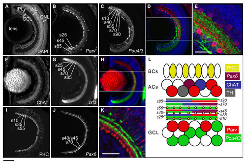
IPL organization of the larval zebrafish retina. Confocal images of horizontal sections of 5 dpf retina stained by immunohistochemistry or with DAPI (nuclear dye). IPL sublaminae are labeled (s10, s25, and so on). (A) DAPI stain shows the basic organization of the retina into GCL, IPL, INL, and ONL. (B-E) Neurites from Parv+ ACs (red) and Pou4f3:mGFP+ GCs (green) are closely apposed but reside in distinct sublaminae. (F-H) Neurites from ChAT+ ACs (red) overlap with zrf3 label (green) in the same sublaminae. (I-K) PKC+ BC axon terminals (red) and Pax6:mGFP+ AC neurites (green) each form three sublaminae that are closely nested but not co-localized. (L) Schematic of IPL organization. Cell types and their neurites are labeled according to the color code on the right. Space is shown between sublaminae for clarity. TH, tyrosine hydroxylase. Bottom left scale bar, for whole retina images, is 50 μm; bottom left scale bar in E, K is 25 μm. |