
Fig. S4
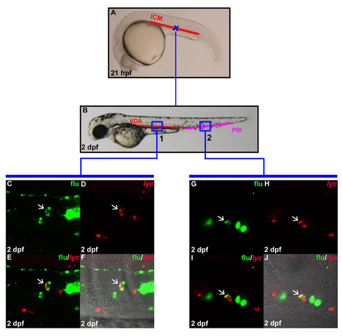
In situ generation of lyc positive myeloid cells in the VDA. (A) Lateral view of 21 hpf embryo indicates the uncaging positions (cross) in the anterior part of the ICM. (B) Lateral view of 2 dpf embryo. The boxed regions show the relative positions in the VDA (region 1) and PBI (region 2) where flu and lyc RNA double-positive cells are found after uncaging. (C,D) Confocal images of the boxed region 1 in B show the flu signal and lyc staining in the VDA at 2 dpf after uncaging at cross A. (E) Merged image of C and D. (F) Superimposed view of E and DIC image. (G,H) Confocal images of the boxed region 2 in B show the flu signal and lyc RNA staining in the PBI at 2 dpf after uncaging at cross in A. (I) Merged image of G and H. (J) Superimposed view of I and DIC image. White arrows indicate the co-staining of flu and lyc RNA. |