
Fig. 1
- ID
- ZDB-FIG-090113-66
- Publication
- Kim et al., 2009 - The extracellular matrix protein TGFBI promotes myofibril bundling and muscle fibre growth in the zebrafish embryo
- Other Figures
- All Figure Page
- Back to All Figure Page
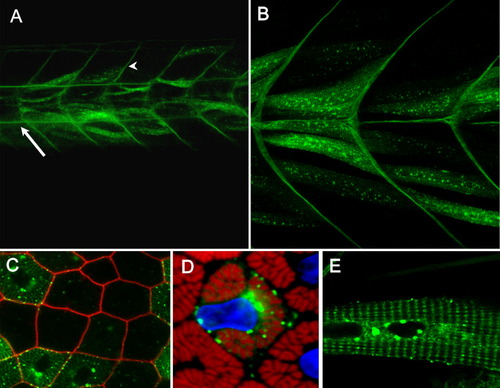
Tgfbi-GFP localises to myosepta and skeletal muscle fibres. Transient mosaic expression of a cDNA encoding a GFP-tagged form of Tgfbi downstream of the α-actin promoter. A,B: The fusion protein accumulated in a striated pattern along individual muscle fibres (shown in detail in E). In addition, protein localised along the notochord (A, arrow) and to the vertical myosepta (arrowhead) at 50 hpf. C: Transverse section of muscle fibres labelled with Lyn-tdTomato (red) reveals its localisation along the sarcolemma and also along the T-tubules that surround myofibrils. D: Perinuclear accumulation of the fusion protein is also apparent (blue: Topro3; red Phalloidin). |