|
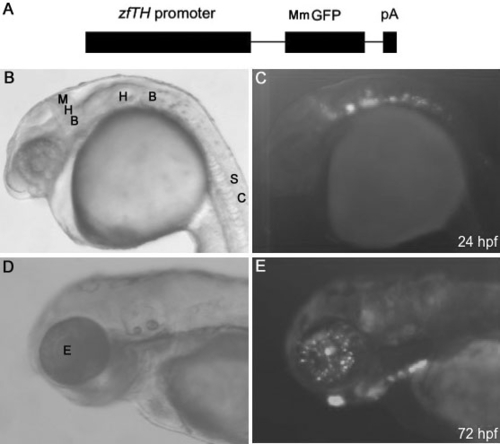
TH promoter-driven GFP expression during zebrafish development. A: Schematic map of the Tg(-12th:MmGFP) construct. The 12 kb sequence containing the zebrafish TH (zfTH) promoter was ligated with MmGFP followed by an SV40 polyadenylation tail (pA). B and C display the bright-field image, and fluorescence image of GFP expression in the embryonic neural system at 24 hpf, respectively. D and E illustrate the bright-field image of the embryonic zebrafish, and fluorescence image of GFP expression in the embryonic retina at 72 hpf, respectively. For B - E, dorsal is on the top and anterior is on the left. Abbreviations: Midbrain and hindbrain boundary (MHB); hindbrain (HB); spinal cord (SC); eye (E).
|