
FIGURE
Fig. 7
- ID
- ZDB-FIG-090112-14
- Publication
- Meng et al., 2008 - Targeting retinal dopaminergic neurons in tyrosine hydroxylase-driven green fluorescent protein transgenic zebrafish
- Other Figures
- All Figure Page
- Back to All Figure Page
Fig. 7
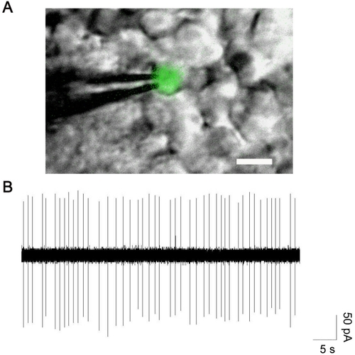
In isolated whole mount retina, GFP-labeled neurons exhibit spontaneous spikes. Merged fluorescence and infrared images of a Tg(-12th:MmGFP) neuron and recording electrode in a whole mount zebrafish retina is shown in panel A. Scale bar equals 10 μm. Panel B displays spontaneous spikes recorded in a GFP-labeled cell. Loose-patch recordings were made using a voltage-clamp mode with an electrode holding potential of 0 mV. |
Expression Data
Expression Detail
Antibody Labeling
Phenotype Data
Phenotype Detail
Acknowledgments
This image is the copyrighted work of the attributed author or publisher, and
ZFIN has permission only to display this image to its users.
Additional permissions should be obtained from the applicable author or publisher of the image.