|
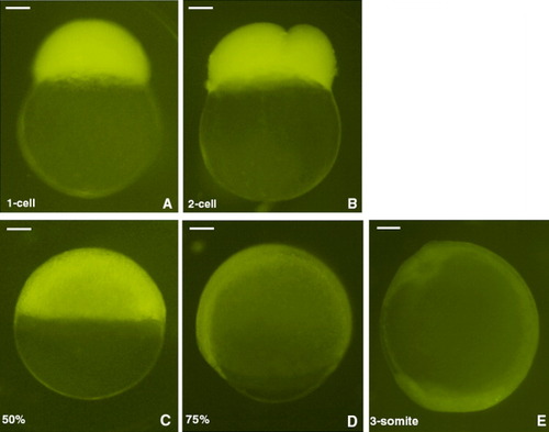
Expression of enhanced yellow fluorescent protein (eYFP) in embryos from a female transgenic zebrafish. Nontransgenic embryos are produced by a female transgenic zebrafish (heterozygous for the transgene) mated with a male wild-type zebrafish. Images are photographed by the DP70 digital camera (Olympus, Japan) with 5-sec exposure time. Embryos are laterally viewed and positioned animal pole top (A-D) or anterior top (E). A: Fertilized egg at one-cell stage. B: Two-cell. C: 50% epiboly. D: 75% epiboly. E: Three-somite stage (11 hours postfertilization [hpf]). eYFP expressions appearing in these stages are due to maternal loading of eYFP protein (A-C) or both eYFP protein and mRNA (D,E). Scale bar = 100 μm.
|