
Fig. 3
- ID
- ZDB-FIG-081205-5
- Publication
- Blin et al., 2008 - NR4A2 controls the differentiation of selective dopaminergic nuclei in the zebrafish brain
- Other Figures
- All Figure Page
- Back to All Figure Page
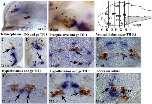
Compared expression of NR4A2b and TH during zebrafish brain development. Localization of NR4A2b transcripts obtained by in situ hybridization (blue staining) and of TH protein obtained by immunochemistry (brown). (A,B) Flat-mounted deyolked whole-mount embryos, anterior to the left, dorsal to the top, (A) lateral view of a 24 hpf embryo, (B) lateral view of a 48 hpf embryo, focus on the posterior tuberculum and hypothalamus. (C–I) Cross sections of brain at 72 hpf, dorsal to the top, in order along the antero-posterior axis. The brain drawing (top right panel) shows the position of the sections. Black arrows indicate cellular co-expression, and the numbers refer to TH cell groups (Rink and Wullimann, 2002). Lower magnifications of these sections are presented in SD13. |
| Genes: | |
|---|---|
| Antibody: | |
| Fish: | |
| Anatomical Terms: | |
| Stage Range: | Prim-5 to Protruding-mouth |
Reprinted from Molecular and cellular neurosciences, 39(4), Blin, M., Norton, W., Bally-Cuif, L., and Vernier, P., NR4A2 controls the differentiation of selective dopaminergic nuclei in the zebrafish brain, 592-604, Copyright (2008) with permission from Elsevier. Full text @ Mol. Cell Neurosci.