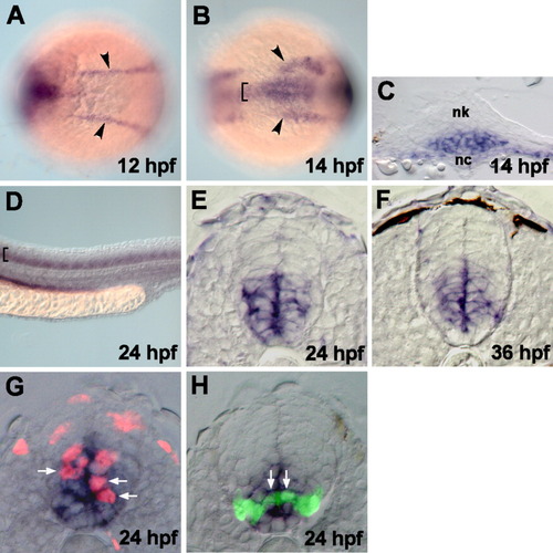
fz8a expression revealed by in situ RNA hybridization. A,B: Dorsal views of the trunk region of whole embryos, anterior to the left. Arrowheads and brackets indicate expressions in the prechordal plate and medial neuroectoderm, respectively. C: Transverse sections, dorsal to the top, through the trunk region. Ventral neural keel (nk) cells, overlying notochord (nc), started to express fz8a at 14 hpf. D: Lateral view of the spinal cord of whole embryos, dorsal to the top and anterior to the left. Brackets indicate the expression of fz8a in the ventral spinal cord cells. E-H: Transverse sections of the spinal cord, dorsal to the top. E, F: fz8a-expressing cells occupy the ventral neural precursor cells. G: fz8a-expressing cells incorporate BrdU, as indicated by anti-BrdU antibody staining (red color), indicating that they are proliferating precursors. Arrows indicate fz8a+, BrdU+ neural precursor cells. H: fz8a-expressing cells confer olig2+ precursors, which are marked by EGFP fluorescence in the Tg(olig2:egfp) embryo. Arrows indicate fz8a+, olig2+ precursor cells in the ventral spinal cord.
|
