
Fig. 3
- ID
- ZDB-FIG-080904-7
- Publication
- Marlow et al., 2008 - Bucky ball functions in Balbiani body assembly and animal-vegetal polarity in the oocyte and follicle cell layer in zebrafish
- Other Figures
- All Figure Page
- Back to All Figure Page
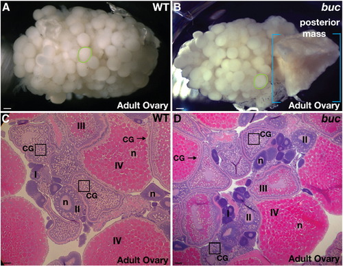
bucky ball ovaries are grossly normal. (A) Wild-type and (B) buc mutant ovaries from adults are of similar size and composition (green circles outline single late stage III or IV oocytes), except that older buc mutant ovaries often have a mass of tissue in the posterior region of the ovary (blue brackets highlight the extent of the posterior mass). Other aspects of oogenesis (oocyte stages I–IV) appeared normal in buc mutants, as revealed by H and E stained adult ovary sections from (C) wild-type and (D) buc. (A, B) Dissecting microscope images. (C, D) 10x images. CG indicates cortical granules located centrally in young oocytes (e.g. box) and at the cortex (arrow) in late stage WT and mutant oocytes. n indicates the oocyte nucleus. Scale bars, 200 μm (A, B); 50 μm (C, D). |
| Fish: | |
|---|---|
| Observed In: | |
| Stage: | Adult |
Reprinted from Developmental Biology, 321(1), Marlow, F.L., and Mullins, M.C., Bucky ball functions in Balbiani body assembly and animal-vegetal polarity in the oocyte and follicle cell layer in zebrafish, 40-50, Copyright (2008) with permission from Elsevier. Full text @ Dev. Biol.