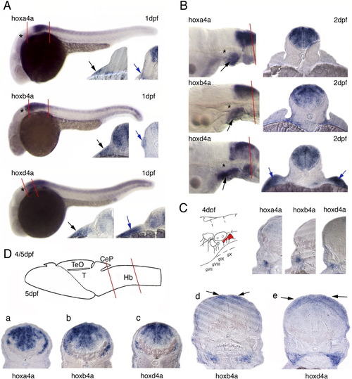
Hoxa4a, hoxb4a, and hoxd4a expression in developing embryos and larvae. A: In situ hybridized embryos at 1 dpf. The otic vesicle is marked by an asterisk. Transverse sections were performed through the anterior r7 and posterior. The levels of sections are indicated. Black arrows indicate signal in the epibranchial placode. Hybridization signals below the placode indicate expression in the branchial arches. Blue arrows in further posterior sections point to (1) signal adjacent to the somitic mesoderm (hoxa4a), (2) signal in lateral line neuromasts (hoxb4a), and (3) expression in the fin anlage (hoxd4a). B: Hox4 expression in the posterior hindbrain and in the branchial arches (black arrows) at 2 dpf with 20μm sections on 3rd somite level. The blue arrows point to signals in the fin (hoxd4a). C: Schematic drawing of the ganglia VII to X and 20μm sections performed through hybridized larvae at 4dpf with mRNA-transcript in the vagal ganglion. D: Schematic drawing of a 5-dpf zebrafish brain modified from Mueller and Wullimann ([2005]) indicating the level of sections through the hindbrain (a-c) and through the cerebellum (d,e). The black arrows point to hybridization signal in the cerebellum. CEP, cerebellar plate; Hb, hindbrain; TeO, tectum opticum; T, midbrain tegmentum.
|
