
Fig. 3
- ID
- ZDB-FIG-080828-19
- Publication
- Goessling et al., 2008 - APC mutant zebrafish uncover a changing temporal requirement for wnt signaling in liver development
- Other Figures
- All Figure Page
- Back to All Figure Page
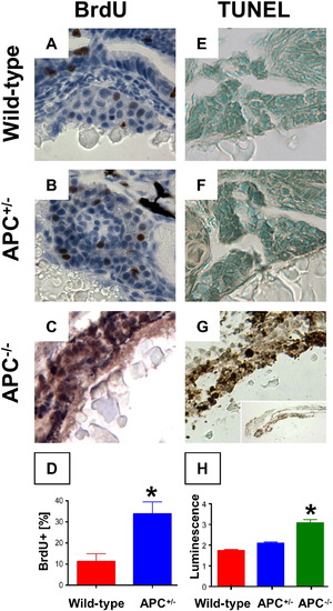
Endodermal proliferation and cell death are dependent on β-catenin levels. (A–D) BrdU incorporation in liver sections corresponding to Figs. 1I, J was significantly upregulated in APC+/- embryos (33.8 ± 12.6% vs. 11.3 ± 7.4%; n = 5, p = 0.016, 40′x), and also in the endodermal region of APC-/- embryos. (E–H) TUNEL staining demonstrated no apoptosis in wild-type and APC+/- embryos, while a high number of TUNEL+ cells were found in the endoderm of APC-/- embryos. (L) Caspase-3 and 7 activity was significantly increased in APC-/- mutants compared to wild-type and APC+/- (ANOVA, n = 10/genotype, p < 0.00001). Significant differences are indicated with an asterisk (∗). |
| Antibody: | |
|---|---|
| Fish: | |
| Condition: | |
| Anatomical Terms: | |
| Stage: | Protruding-mouth |
| Fish: | |
|---|---|
| Condition: | |
| Observed In: | |
| Stage: | Protruding-mouth |
Reprinted from Developmental Biology, 320(1), Goessling, W., North, T.E., Lord, A.M., Ceol, C., Lee, S., Weidinger, G., Bourque, C., Strijbosch, R., Haramis, A.P., Puder, M., Clevers, H., Moon, R.T., and Zon, L.I., APC mutant zebrafish uncover a changing temporal requirement for wnt signaling in liver development, 161-174, Copyright (2008) with permission from Elsevier. Full text @ Dev. Biol.