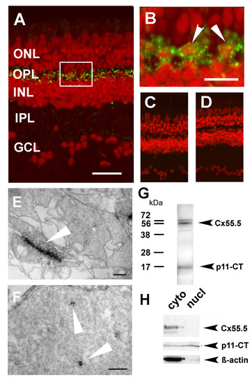
Localization of Cx55.5 immunoreactivity in the nucleus of horizontal cells. A) Confocal imaging using z-stack recordings reproducibly showed that Cx55.5 (green fluorescence) was exclusively expressed in horizontal cells of the outer retina. Scale bar: 100 μm B) The higher magnification of the region of interest (frame in A) provided evidence for small clusters of Cx55.5 immunoreactivity inside the nuclei of HCs (see arrowheads). Scale bar: 20 μm. Antibody controls, C) omitting the primary antibody and D) blocking of the primary antibody by pre-absorption with 1 μg GST-Cx55.5 fusion protein. Nuclei were stained with Sytox Orange (Invitrogen, Karlsruhe, Germany). In order to exclude the possibility that false positive fluorescent emission was recorded from out of focus planes electron microscopical immunohistochemistry of adult retina was performed. Gap junction plaques (arrow) between HCs as shown in E) resemble characteristic membrane associated structures detected with the Cx55.5 antibody, whereas the formation of small clusters (arrows) within the nucleus as shown in F) was typical for nuclear Cx55.5 immunoreactivity. The localization and number of clusters in a single section as shown in the inset was variable but did not exceed N = 3. Note that Cx55.5 immunoreactivity was never found outside the HC layer. Scale bars: 400 nm (E), 300 nm (F). (G) Western blot analysis of total protein extracts isolated from adult retina indicates a doublet of bands at the position 56 kDa and a low molecular weight band at the position 16 kDa. H) Western blot analysis of protein extracts isolated by subcellular fractionation of the adult retina (cyto = cytosolic fraction, nucl = nuclear fraction). The full length Cx55.5 protein product was enriched in the cytoplasmic fraction which is composed of soluble cytosolic proteins and membrane proteins (upper lanes). The 16 kDa protein product was found in the cytosolic and nuclear fraction (middle lanes). The lower blot depicts the β-actin control. Note the faint β-actin band in the nuclear fraction indicating minimal contamination with cytoplasmic remnants. The results shown derived from two independent western blot runs using the same samples (upper and lower lanes: 10% SDS-PAGE; middle lane; 15% SDS-PAGE).
|
