
Fig. 5
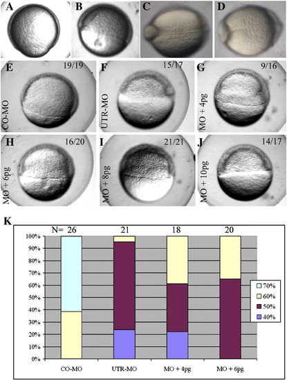
Impaired development resulting from pcdh18a knockdown, and rescue by pcdh18a RNA co-injection. CO-MO (A, C) or pcdh18a ATG-MO (5 ng) (B, D) were injected into embryos at the one-cell stage. ATG-MO-injected embryos (B) showed delayed epiboly (11 of 17, 74%, in one of five experiments) compared to controls (A). Likewise, as control MO-injected embryos reached the 5 somite stage (C), ATG-MO-injected embryos contained 3–4 somites that were wider than normal (D; 25 of 32, 78%). (E–J) Phenotype caused by UTR-MO and its rescue. UTR-MO (F–J) was injected alone (F) or together with 4, 6, 8, and 10 pg pcdh18a RNA (G–J, respectively); CO-MO is in panel E. The UTR-MO-injected embryos showed a delay in epiboly that was rescued by co-injection of the RNA. Quantification of one of four similar experiments is shown in panel K. The color code refers to the percentage of embryos in the respective stage of epiboly at the same time point. |
| Fish: | |
|---|---|
| Knockdown Reagents: | |
| Observed In: | |
| Stage Range: | Shield to 5-9 somites |
Reprinted from Developmental Biology, 318(2), Aamar, E., and Dawid, I.B., Protocadherin-18a has a role in cell adhesion, behavior and migration in zebrafish development, 335-346, Copyright (2008) with permission from Elsevier. Full text @ Dev. Biol.