
Fig. 4
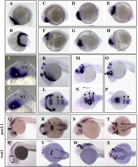
Hatching gland mislocalization in Pcdh18a-overexpressing embryos. In situ hybridization with hgg1 on 12-somite stage embryos (A–J) injected with 100 pg GFP RNA (A, B) or 100 pg pcdh18a RNA (C–J). In two experiments, 13 of 24 (54%) of the pcdh18a RNA-injected embryos showed mislocalization of hgg1, while none of the GFP-injected embryos (n = 18) did. (I, J) show two examples of hgg1 staining (asterisk) in relation to the neural tube (NT), taken at higher magnification in anterior view. (K–P) In situ hybridization with hgg1 (asterisk), krox20 (arrow) and eng2 (arrowhead) at 24hpf, shows mislocalization of hgg1-expressing cells, but an almost normal pattern of krox20 and eng2 in embryos injected with 100 pg pcdh18a RNA (37 of 50, 74%, in three experiments; M–P). None of the 34 GFP RNA-injected embryos showed this phenotype (K, L). (Q–X) Neural tube duplications in pcdh18a RNA-injected embryos at 28hpf were visualized by in situ hybridization with pax2.1 (S, T), and wnt1 (W, X); control embryos are shown in panels Q, R, U, V. Combining data from several experiments using different markers showed duplications in 119 of 202 (59%) injected embryos. |
Reprinted from Developmental Biology, 318(2), Aamar, E., and Dawid, I.B., Protocadherin-18a has a role in cell adhesion, behavior and migration in zebrafish development, 335-346, Copyright (2008) with permission from Elsevier. Full text @ Dev. Biol.