
FIGURE
Fig. 4
- ID
- ZDB-FIG-080613-28
- Publication
- Langenberg et al., 2008 - The eye organizes neural crest cell migration
- Other Figures
- All Figure Page
- Back to All Figure Page
Fig. 4
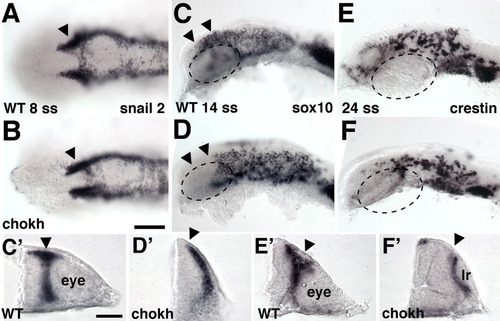
Neural crest cells (NCCs) in chokh embryos. A-F: Wild-type (WT) and chokh mutant embryos labeled with in situ hybridization for NC markers at the indicated stages. Arrowheads mark normal (A-C,C′,E′) and missing (D,D′,F′) NCC populations. The hatched circle marks the eye in WT embryos (C,E) and the approximate position where the eye would normally have developed in chokh embryos (D,F). A, B are dorsal, C-F lateral views, anterior to the left. C′-F′: Cross-sections through the eye vesicle in WT and the respective region in chokh embryos. Scale bar = 100 μm in A-F, 50 μm in C′-F′. lr, lens remnant. |
Expression Data
| Genes: | |
|---|---|
| Fish: | |
| Anatomical Term: | |
| Stage Range: | 5-9 somites to 20-25 somites |
Expression Detail
Antibody Labeling
Phenotype Data
Phenotype Detail
Acknowledgments
This image is the copyrighted work of the attributed author or publisher, and
ZFIN has permission only to display this image to its users.
Additional permissions should be obtained from the applicable author or publisher of the image.
Full text @ Dev. Dyn.