FIGURE

Fig. S7

ID
ZDB-FIG-080605-7
Publication
Sidi et al., 2008 - Chk1 Suppresses a Caspase-2 Apoptotic Response to DNA Damage that Bypasses p53, Bcl-2, and Caspase-3
Other Figures
All Figure Page
Back to All Figure Page
Fig. S7

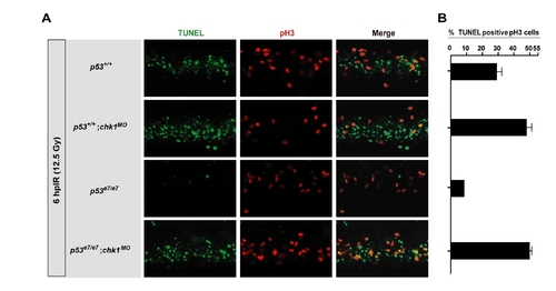
Chk1-suppressed apoptosis occurs predominantly during the interphase in zebrafish embryos
(A) Confocal images of the caudal spinal cord in representative embryos of each indicated genotype processed for TUNEL (green)/pH3 (red) double-labeling. While approximately half of the pH3-labeled cells in irradiated p53e7/e7;chk1MO embryos are TUNEL positive (see panel B), the bulk of TUNEL-stained nuclei are pH3 negative, implying that death mainly occurs during the interphase. By contrast, cisplatin- or doxorubicin-induced death of MK-2–depleted Tp53-/- MEFs occurs exclusively in mitosis (Reinhardt et al., 2007).
(B) Percentage of double-labeled cells among >200 pH3-positive cells visualized on images as in panel A. Data are means ± SEM.

Expression Data

Expression Detail
Antibody Labeling
Phenotype Data

Phenotype Detail
Acknowledgments
This image is the copyrighted work of the attributed author or publisher, and ZFIN has permission only to display this image to its users. Additional permissions should be obtained from the applicable author or publisher of the image.

Reprinted from Cell, 133(5), Sidi, S., Sanda, T., Kennedy, R.D., Hagen, A.T., Jette, C.A., Hoffmans, R., Pascual, J., Imamura, S., Kishi, S., Amatruda, J.F., Kanki, J.P., Green, D.R., D'Andrea, A.A., and Look, A.T., Chk1 Suppresses a Caspase-2 Apoptotic Response to DNA Damage that Bypasses p53, Bcl-2, and Caspase-3, 864-877, Copyright (2008) with permission from Elsevier. Full text @ Cell