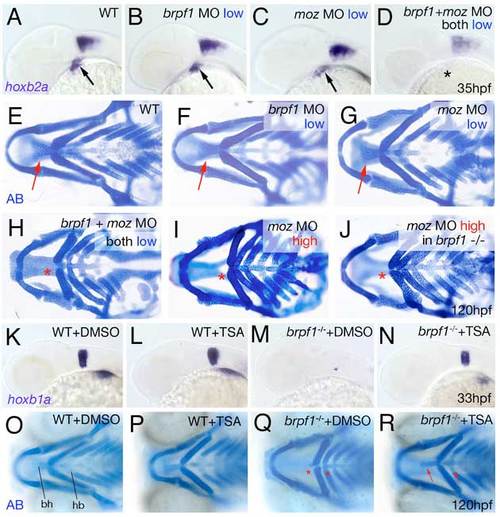
Brpf1 genetically interacts with Moz and brpf1 mutant defects are alleviated by HDAC inactivation with TSA. (A-D) hoxb2a in situ hybridizations at 35 hpf, lateral views. Arrows point to hoxb2a expression in cranial neural crest of wild-type embryo (A) and embryos injected with low amounts of brpf1 MO (B) or moz MO (C); asterisk indicates absent hoxb2a expression in embryo co-injected with the same low amounts of both MOs (D). (E-J) Alcian Blue staining of head skeletons at 120 hpf, ventral view. Arrows point to basihyal in wild-type (E), and larvae injected with low amounts of brpf1 MO (F) or moz MO (G). Asterisks indicate absent basihyal in larva co-injected with low amounts of brpf1 and moz MO (H), in strong moz morphant (I) and in brpf1 mutant injected with high amounts of moz MO (J). Co-injection of low doses of brpf1 MO and moz MO, which in single injections did not cause any phenotypes (compare F and G with E), caused defects as severe as in strongest brpf1 or moz morphants (compare H with I; n=11/13). By contrast, brpf1 mutants injected with highest amounts moz MOs showed a phenotype no more severe than that of moz single morphants (compare J with I; n=20/20). (K-N) TSA treatment enhances hoxb1a expression in r4 of both wild-type and brpf1 mutant embryos. hoxb1a in situ hybridizations at 33 hpf, lateral view of head region. TSA-treated wild-type embryo (L) displays slightly stronger hoxb1a expression than DMSO-treated sibling (K; n=27/35). TSA-treated brpf1 mutant displays recovered hoxb1a expression in r4 (N; embryo genotyped after photography; n=11/11), whereas expression remains absent in control mutant treated with DMSO (M). (O-R) TSA treatment from 20-33 hpf significantly ameliorates the later cartilage defects of brpf1 mutants. Alcian Blue staining of head skeletons at 120 hpf, ventral views. In O, basihyal of arch 2 (bh) and hypobranchials (hb) of arch 3 are indicated. The head skeleton of the TSA-treated wild-type larva (P) looks similar to that of the DMSO-treated control sibling (O). The TSA-treated brpf1 mutant (R) displays a significant amelioration of skeletal defects, including formation of hypobranchials and a normally sized basihyal (red arrows; animal genotyped after photography; n=26/33). By contrast, both elements remain absent or strongly reduced in control mutant treated with DMSO (Q; red asterisks; n=9/9). Stronger or longer TSA treatments interfered with additional developmental processes requiring HDAC activity and caused defects comparable to those of zebrafish hdac1 mutants (Pillai et al., 2004), including more severe craniofacial abnormalities, which masked the brpf1-specific traits.
|
