
Fig. 2
- ID
- ZDB-FIG-080529-45
- Publication
- Rawls et al., 2004 - Gnotobiotic zebrafish reveal evolutionarily conserved responses to the gut microbiota
- Other Figures
- All Figure Page
- Back to All Figure Page
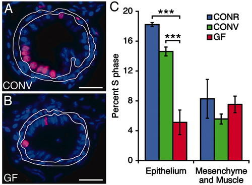
The microbiota stimulates intestinal epithelial proliferation. (A and B) Sections prepared from the intestines of 6-dpf CONV and GF zebrafish after a 24-h exposure to bromodeoxyuridine in their environmental water. Sections were stained with antibodies to bromodeoxyuridine (magenta) and the nuclear stain bisbenzimide (blue). The mesenchyme and muscle surrounding the intestinal epithelium are outlined in white. (C) Quantitation of S-phase cells in the intestinal epithelium and mesenchyme. The percentage of cells in S phase in GF intestinal epithelium is significantly lower than in CONR or CONV animals (P < 0.0001, indicated by brackets with three asterisks). Data are expressed as the mean of two independent experiments ± SEM (n = 19-31 sections scored per animal; ≥6 animals per experiment). (Bars: 25 μm in A and B.) |