
FIGURE
Fig. 3
- ID
- ZDB-FIG-080514-16
- Publication
- Lister et al., 2001 - Duplicate mitf genes in zebrafish: complementary expression and conservation of melanogenic potential
- Other Figures
- All Figure Page
- Back to All Figure Page
Fig. 3
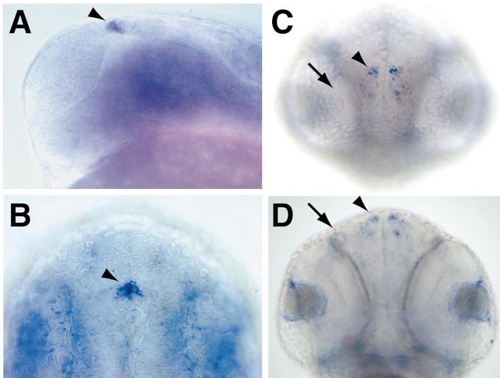
Expression of mitfb in nonpigment cells. (A) 24 hpf, lateral view; (B) dorsal view. Expression of mitfb in the epiphysis (arrowheads). (C) 48 hpf, frontal view; (D) 48 hpf, ventral view. mitfb is detected in symmetrical clusters of cells in the olfactory bulbs (arrowheads; arrows indicate olfactory organ). |
Expression Data
| Gene: | |
|---|---|
| Fish: | |
| Anatomical Terms: | |
| Stage Range: | Prim-5 to Long-pec |
Expression Detail
Antibody Labeling
Phenotype Data
Phenotype Detail
Acknowledgments
This image is the copyrighted work of the attributed author or publisher, and
ZFIN has permission only to display this image to its users.
Additional permissions should be obtained from the applicable author or publisher of the image.
Reprinted from Developmental Biology, 237(2), Lister, J., Close, J., and Raible, D., Duplicate mitf genes in zebrafish: complementary expression and conservation of melanogenic potential, 333-344, Copyright (2001) with permission from Elsevier. Full text @ Dev. Biol.