
FIGURE
Fig. 4
- ID
- ZDB-FIG-080513-13
- Publication
- Herbomel et al., 2001 - Zebrafish early macrophages colonize cephalic mesenchyme and developing brain, retina, and epidermis through a M-CSF receptor-dependent invasive process
- Other Figures
- All Figure Page
- Back to All Figure Page
Fig. 4
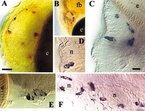
Early macrophages around olfactory organs and neuromasts. (A, B) Neutral red vital staining of macrophages associated with the left olfactory organ at 50 (A) and 84 hpf (B, low magnification). (C–F) In situ hybridization for L-plastin; (C) Right olfactory organ, 96 hpf, dorsal view. (D) Two macrophages, probably sister cells from a recent mitosis, adjacent to an anterior neuromast at 120 hpf. (E) A latero-ventral neuromast just caudal to the eye at 120 hpf. (F) Two neuromasts of the supra-orbital line at 144 hpf. (fb) forebrain; (e) eye; (n) neuromast. Bars: (A) 20 μm; (C–F) 10 μm. |
Expression Data
Expression Detail
Antibody Labeling
Phenotype Data
Phenotype Detail
Acknowledgments
This image is the copyrighted work of the attributed author or publisher, and
ZFIN has permission only to display this image to its users.
Additional permissions should be obtained from the applicable author or publisher of the image.
Reprinted from Developmental Biology, 238(2), Herbomel, P., Thisse, B., and Thisse, C., Zebrafish early macrophages colonize cephalic mesenchyme and developing brain, retina, and epidermis through a M-CSF receptor-dependent invasive process, 274-288, Copyright (2001) with permission from Elsevier. Full text @ Dev. Biol.