
Fig. 1
- ID
- ZDB-FIG-080506-10
- Publication
- Biemar et al., 2001 - Pancreas development in zebrafish: early dispersed appearance of endocrine hormone expressing cells and their convergence to form the definitive islet
- Other Figures
- All Figure Page
- Back to All Figure Page
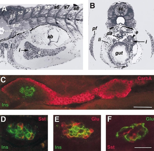
Morphology of the exocrine and endocrine pancreas in the zebrafish larva. (A) Sagittal and (B) cross sections of 6-day-old larvae showing the overall organization of the endocrine (i, arrow) and exocrine pancreatic tissues (e in A, and e, arrowhead in B). (C–F) Confocal images of pancreatic islet tissue showing whole-mount immunofluorescence staining performed on (C) 5.5-day-old, (D) 4-days, and (E, F) 3-day-old embryos with a combination of (C) anti-carboxypeptidase A and anti-insulin; (D) anti-insulin and anti-somatostatin; (E) anti-insulin and anti-glucagon; (F) anti-somatostatin and anti-glucagon antisera. In A and C, anterior is oriented to the left. da, dorsal aorta; e; exocrine tissue; i, islet; li, liver; o, otolith; pf, pectoral fin; sb, swim bladder; sc, spinal cord; s1–8, somite 1 to 8. Scale bar, 50 μm in C and 25 μm in F (same magnification as D and E). |
| Gene: | |
|---|---|
| Antibodies: | |
| Fish: | |
| Anatomical Terms: | |
| Stage Range: | Protruding-mouth to Day 5 |
Reprinted from Developmental Biology, 230(2), Biemar, F., Argenton, F., Schmidtke, R., Epperlein, S., Peers, B., and Driever, W., Pancreas development in zebrafish: early dispersed appearance of endocrine hormone expressing cells and their convergence to form the definitive islet, 189-203, Copyright (2001) with permission from Elsevier. Full text @ Dev. Biol.