
Fig. 6
- ID
- ZDB-FIG-080423-31
- Publication
- Riley et al., 2003 - Ringing in the new ear: resolution of cell interactions in otic development
- Other Figures
- All Figure Page
- Back to All Figure Page
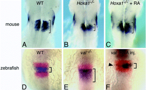
Expression of Fgf3 at comparable stages in mouse and zebrafish. Brackets are shown in all panels to help gauge the size of the Fgf3 domain in the hindbrain. (A–C) Mouse embryos at embryonic day 8.5. (A) A wild-type embryo showing expression in r5 and r6. (B) A Hoxa1-/- mutant shows a greatly reduced hindbrain domain. (C) Brief treatment of Hoxa1-/- mutants with RA restores Fgf3 expression to normal [Pasqualetti et al 2001]. (D–F) Zebrafish embryos at the six-somite stage showing expression of fgf3 (blue) and krox20 (red). (D) Wild-type embryos express fgf3 in r4 and krox20 in r3 and r5. (E) val-/- mutants show loss of krox20 in r5 and expansion of fgf3 into the r5/6 region. (F) A wild-type embryo injected with val mRNA at the 2 cell stage. Expression is primarily restricted to the left side of the embryo where hindbrain expression of fgf3 is nearly extinguished (arrowhead). Expression on the right is essentially normal. Expression of fgf8 in r4 is not altered by either loss of val or misexpression of val [Kwak et al 2002]. With permission, (A–C) are reprinted from [Pasqualetti et al 2001], and (D–F) are reprinted from [Kwak et al 2002]. |
Reprinted from Developmental Biology, 261(2), Riley, B.B. and Phillips, B.T., Ringing in the new ear: resolution of cell interactions in otic development, 289-312, Copyright (2003) with permission from Elsevier. Full text @ Dev. Biol.