
Fig. 5
- ID
- ZDB-FIG-080423-14
- Publication
- Parichy et al., 2003 - Zebrafish puma mutant decouples pigment pattern and somatic metamorphosis
- Other Figures
- All Figure Page
- Back to All Figure Page
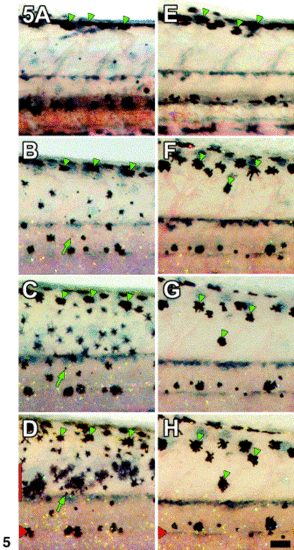
EL melanophore movements and appearance of metamorphic melanophores differs between wild-type and puma mutant larvae. (A–D) In wild-type larvae, most melanophores comprising the dorsal stripe of the embryonic/early larval pigment pattern remain near the dorsal aspect of the myotomes during early metamorphosis (arrowheads show representative cells). Additional newly differentiated melanophores appear as initially smaller, more lightly melanized cells scattered over the flank. Arrow in B–D, a newly differentiated melanophore moving dorsally to join the dorsal stripe. A–D, 15, 16, 19, 21 dpf, respectively. Red bar in (H), region of developing dorsal primary melanophore stripe. (E–H) In puma mutants, early larval dorsal stripe melanophores migrate further ventrally toward the middle of the flank than in wild-type (arrowheads). Newly differentiated melanophores are not evident. E–H, 15, 18, 19, 21 dpf, respectively. Images are scaled to maintain the same relative anterior–posterior positions of cells. Red arrowheads in (D) and (H), horizontal myosepta. Scale bar, 100 μm (H only). |
Reprinted from Developmental Biology, 256(2), Parichy, D.M. and Turner, J.M., Zebrafish puma mutant decouples pigment pattern and somatic metamorphosis, 242-257, Copyright (2003) with permission from Elsevier. Full text @ Dev. Biol.