
- Title
-
Zebrafish puma mutant decouples pigment pattern and somatic metamorphosis
- Authors
- Parichy, D.M. and Turner, J.M.
- Source
- Full text @ Dev. Biol.
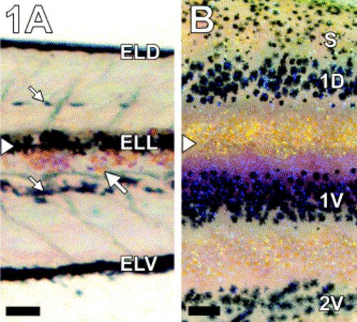
Larval-to-adult transformation of the zebrafish pigment pattern. (A) Early larval pigment pattern. Shown is an individual at 14 dpf, at the onset of pigment pattern metamorphosis. Established during embryogenesis, the early larval pattern of melanophore stripes persists through the onset of metamorphosis. On the trunk, this pattern consists of an early larval dorsal melanophore stripe (ELD), an early larval lateral melanophore stripe (ELL) along the horizontal myoseptum, and an early larval ventral melanophore stripe (ELV). Below the plane of focus, melanophores also can be seen along the dorsal neural tube and the dorsal aorta (small arrows). At the onset of pigment pattern metamorphosis, a few xanthophores and iridophores (large arrow) are apparent immediately ventral to the early larval lateral melanophore stripe. (B) An early adult/juvenile pigment pattern develops by terminal stages of pigment pattern metamorphosis. Shown is an individual at 37 dpf. Two adult primary melanophore stripes have developed dorsal (1D) and ventral (1V) to a light interstripe region containing xanthophores and iridophores. The first adult secondary melanophore stripe (2V) has started to form near the base of the anal fin. Dorsally, melanophores and xanthophores are scattered over the myotomes and populate the dorsal scales (S). Arrowheads in (A) and (B) show location of the horizontal myoseptum; note that EL melanophores initially present at this location (A, ELL) are absent by terminal stages of pigment pattern metamorphosis (B). Images are not to the same scale: Scale bars, (A) 150 μm, (B) 500 μm. |
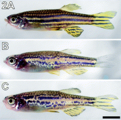
puma mutant adults exhibit disrupted stripes. (A) Wild-type zebrafish exhibit four to five regular dark horizontal stripes with lighter interstripe regions. (B, C) puma mutant adults exhibit fewer stripes with irregular borders. Two examples are shown. Scale bar, 5 mm. |
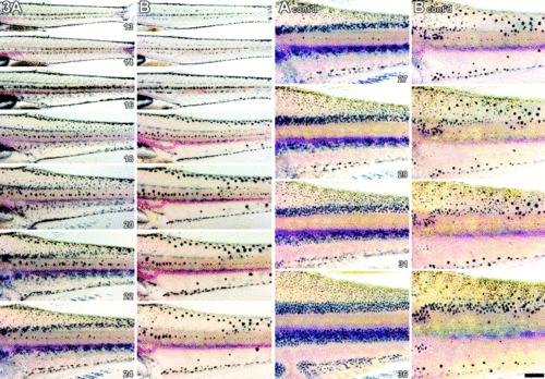
Pigment pattern metamorphosis in wild-type and puma mutant zebrafish. Numbers indicate days postfertilization. (A) Wild-type zebrafish exhibit a gradual increase in melanophore numbers and a gradual emergence of the adult pattern of dark primary and secondary melanophore stripes with light interstripe regions. (B) From embryonic to early larval stages (≤13 dpf), puma mutants exhibit pigment patterns indistinguishable from wild-type. Subsequently, however, puma mutants have fewer melanophores than wild-type fish during pigment pattern metamorphosis and juvenile development. Although some additional melanophores are present on the flank during middle stages of pigment pattern metamorphosis (e.g., 22 dpf), these are much less numerous than in wild-type larvae at corresponding stages. Finally, during terminal stages of pigment pattern metamorphosis and juvenile development (e.g., ≥27 dpf), more melanophores are present, and an irregular striped pattern forms that is nevertheless patchier than in wild-type fish. Scale bar, 500 μm. |
EL melanophore movements and appearance of metamorphic melanophores differs between wild-type and puma mutant larvae. (A–D) In wild-type larvae, most melanophores comprising the dorsal stripe of the embryonic/early larval pigment pattern remain near the dorsal aspect of the myotomes during early metamorphosis (arrowheads show representative cells). Additional newly differentiated melanophores appear as initially smaller, more lightly melanized cells scattered over the flank. Arrow in B–D, a newly differentiated melanophore moving dorsally to join the dorsal stripe. A–D, 15, 16, 19, 21 dpf, respectively. Red bar in (H), region of developing dorsal primary melanophore stripe. (E–H) In puma mutants, early larval dorsal stripe melanophores migrate further ventrally toward the middle of the flank than in wild-type (arrowheads). Newly differentiated melanophores are not evident. E–H, 15, 18, 19, 21 dpf, respectively. Images are scaled to maintain the same relative anterior–posterior positions of cells. Red arrowheads in (D) and (H), horizontal myosepta. Scale bar, 100 μm (H only). |
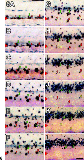
Movement or death of EL lateral stripe melanophores in wild-type larvae. Some EL melanophores initially comprising the lateral stripe move short distances dorsally to join the developing adult dorsal primary melanophore stripe (e.g., melanophores 2, 3). Early larval melanophores that fail to leave this region typically are lost (e.g., melanophores 1, 4, 5; last observed position in yellow). A–L, 14, 15, 16, 17, 18, 19, 20, 21, 23, 25, 26, 29 dpf, respectively. Images are scaled to maintain the same relative anterior–posterior positions of cells. Scale bar, 200 μm (L only). |
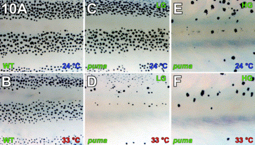
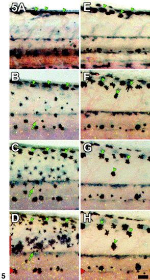
Severity of the puma mutant phenotype depends on temperature and growth rate. (A) Wild-type individuals reared at 24°C develop typical wild-type melanophore stripes. (B) Wild-type individuals reared at 33°C also develop typical wild-type stripes. (C, D) The puma mutant phenotype is temperature-dependent. (C) At 24°C, melanophore densities and patterns approach that seen in wild-type individuals. (D) At 33°C, however, a severe melanophore deficiency and pattern defect is observed. (C–F) The severity of the puma mutant phenotype depends on individual growth rate as well as temperature. LG, low growth. HG, high growth. (C, D) puma mutants reared under conditions favoring a low growth rate (LG) exhibit markedly different phenotypes depending on the temperature they experience during development. (E, F) puma mutants reared under conditions favoring a high growth rate (HG) exhibit similar phenotypes across temperatures, since even individuals reared at 24°C exhibit a severe melanophore deficiency. In contrast to puma mutants, phenotypes of wild-type fish did not differ dramatically given different growth rates (e.g., A, high growth; B, low growth). In all images, the region shown is immediately dorsal to the anal fin. |
Reprinted from Developmental Biology, 256(2), Parichy, D.M. and Turner, J.M., Zebrafish puma mutant decouples pigment pattern and somatic metamorphosis, 242-257, Copyright (2003) with permission from Elsevier. Full text @ Dev. Biol.