
Fig. 2
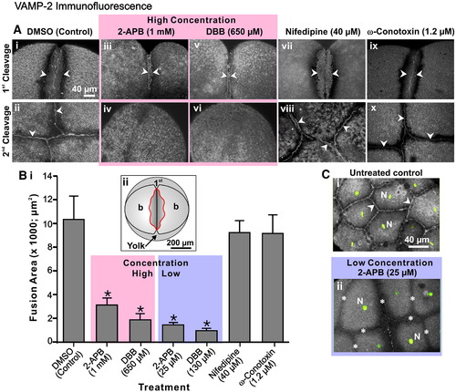
VAMP-2 vesicle fusion requires an elevation in Ca2+ derived from intracellular stores. (A) Embryos were treated after furrow initiation with 2-APB, DBB, nifedipine or ω-conotoxin at the concentrations indicated. They were then fixed during late deepening of the 1st cleavage (see panels Ai, Aiii, Av, Avii and Aix) and at the completion of the 2nd cleavage or 1st furrow regression (see panels Aii, Aiv, Avi, Aviii and Ax) for VAMP-2 immunofluorescence analysis. The regions of VAMP-2 labeling are indicated with arrowheads. Panels Aii, Aviii and Ax indicate the extent of VAMP-2 labeling at the completion of 2nd furrow apposition, whereas panels Aiv and Avi clearly indicate the complete regression of the 1st cleavage furrow and the absence of VAMP-2 labeling in this region in embryos treated with either 2-APB (i.e., 1 mM) or DBB (i.e., 650 μM). (B) The extent of VAMP-2 vesicle fusion during late furrow deepening was quantified at the furrow region by measuring the area of VAMP-2 membrane labeling, indicated in the region within the red line in the schematic shown in panel Bii. Data presented are averaged values ± S.E.M. where n ≥ 6. One-way ANOVA was performed for statistical analysis. The Neuman–Keuls multiple comparison test was performed as a post-hoc analysis. * indicates values significantly different from the DMSO control (at p < 0.001). (C) Untreated control and 2-APB-treated embryos were fixed at the 8- to 16-cell stages for VAMP-2 immunofluorescence (grey-scale). The nuclei (N) were stained with Sytox green (indicated in green). Panels Ci and Cii are 2-D reconstructions from stacks of images captured from an animal pole view. In panel Ci, arrowheads indicate normal VAMP-2 labeling between the fully apposed daughter cells in a control embryo, while in panel Cii the asterisks indicate cleavage furrows in the 2-APB-treated embryos that failed to deepen. |
| Gene: | |
|---|---|
| Fish: | |
| Conditions: | |
| Anatomical Term: | |
| Stage Range: | 1-cell to 2-cell |
Reprinted from Developmental Biology, 316(2), Li, W.M., Webb, S.E., Chan, C.M., and Miller, A.L., Multiple roles of the furrow deepening Ca2+ transient during cytokinesis in zebrafish embryos, 228-248, Copyright (2008) with permission from Elsevier. Full text @ Dev. Biol.