
Fig. 4
- ID
- ZDB-FIG-080411-12
- Publication
- Chen et al., 2008 - Liver-specific expression of p53-negative regulator mdm2 leads to growth retardation and fragile liver in zebrafish
- Other Figures
- All Figure Page
- Back to All Figure Page
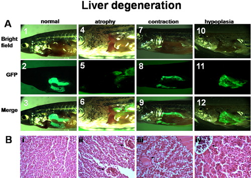
Liver degeneration in Gmdm2-liver fish. A: Whole-body image of normal, atrophic, contractive, and hypoplastic livers in a G-liver and three Gmdm2-liver#1 fish with various levels of growth retardation or illness at 5 months of age. Normal liver (1-3); atrophic liver (4-6); contractive liver (7-9); hypoplastic liver (10-12). Bright field image of the livers (1, 4, 7, and 10); GFP fluorescent image of the same livers (2, 5, 8, and 11); merge image of the same livers (3, 6, 9, and 12). B: Hepatic lesions in Gmdm2-liver transgenic zebrafish. GFP::Mdm2 over-expression leads to progression from normal liver (i) to liver atrophy (ii), contraction (iii), and hyperplasia (iv). Coalescence of hepatic foci as depicted by the arrow. Hematoxylin and eosin (H&E)-stained slides of paraffin-embedded fixed livers from G-liver#1 (1) and from Gmdm2-liver#1 transgenic zebrafish (4, 7, and 10). Magnifications at 100x. |