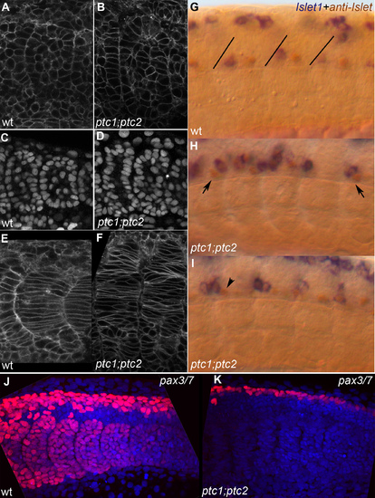
A,C,E,G,J) wild type embryo; B,D,F,H,I,K) ptc1;ptc2 mutants. A,B) lynGFP membrane label showing most recent fully formed somite, 14 somite stage. Double mutant somites still form an epithelium but irregularities in the epithelial somite are more frequent. C,D) DAPI nuclear stain showing essentially the same result as A,B. E,F) LynGFP labeling outlining cells of differentiating somites in a 14 somite wild-type and ptc1;ptc2 mutant embryo, medial optical section through somite 10 and 9+10, respectively. Somites have lost their clear V shape and the number of elongated adaxial cells appears increased. G,H,I) Double labeling showing anti-Islet (brown) and islet1 expression (20 ss). Brown cells express only Islet 2 and are Caudal Primary (CaP) neurons, blue/brown cells express islet 1 (and possibly 2) and are Middle Primary neurons (MiP). In wild-type (G) brown CaPs are located in the middle of each segment whereas the blue/brown MiPs are close to the somite boundary (drawn-in in G for clarity) [54]. In a ptc1;ptc2 mutant background (H,I) mistakes in this order are very frequent, for instance, blue and brown cells close to the posterior of the segment, or the exact mirror image of that (H, arrows). In (I) a brown cell can be seen in the position where a blue cell would be expected (arrowhead). J,K) Z-projection of Pax3/7 labeling (red) and nuclear DAPI stain (blue) on posterior 5–6 somites of 20 somite stage embryos showing the loss of all pax3/7 staining in the somites, and a reduction of the number of pax3/7 positive nuclei in the dorsal neural tube in ptc1;ptc2 double mutants.
|
