
Fig. 3
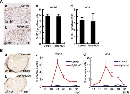
Comparison of cell proliferation and cell death patterns in the lens and retina of control and Fgf19 MO1-injected embryos. A: Control and Fgf19 MO1-injected embryos were stained using an anti-H3P antibody. (a, b) Panels show representative transverse sections of the eye at 22 hpf. (c, d) The percentage of proliferating cells labeled with anti-phosphorylated Histone H3 antibody in the retina and lens of control and Fgf19 MO1-injected embryos. In the lens and retina, the injection of Fgf19 MO1 did not affect cell proliferation. B: Apoptotic cells in the eye of control and Fgf19 MO1-injected embryos were marked by TUNEL staining. (a, b) Panels show their representative transverse sections of the eye at 24 hpf. (c, d) The percentage of apoptotic cells in the retina and lens of control and Fgf19 MO1-injected embryos at 14, 18, 24, 36, 48 and 72 hpf. The ratio of the number of apoptotic cells to the total number of cells in the lens and retina increased significantly in Fgf19 MO1-injected embryos at 24 hpf. |
| Fish: | |
|---|---|
| Knockdown Reagent: | |
| Observed In: | |
| Stage: | Prim-5 |
Reprinted from Developmental Biology, 313(2), Nakayama, Y., Miyake, A., Nakagawa, Y., Mido, T., Yoshikawa, M., Konishi, M., and Itoh, N., Fgf19 is required for zebrafish lens and retina development, 752-766, Copyright (2008) with permission from Elsevier. Full text @ Dev. Biol.