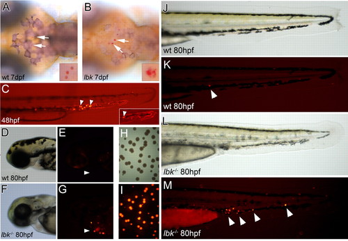
lbk larvae show a compromised ability to clear bacterial infections. (A,B) Dorsal views of larvae in which macrophages were stained with Neutral Red to reveal intracellular vesicles (arrows). Compared with the vesicles observed in wild-type macrophages, those in lbk appear enlarged, heterogeneous in size and less regularly shaped. Insets show higher magnifications of macrophage vesicles. (C) Combined bright-field and fluorescence images of the tail of a 48 hpf larva 5 minutes after the injection of DsRed-fluorescent bacteria, showing bacteria still circulating in the bloodstream (inset; fluorescent traces of bacteria moving in blood vessels) and bacteria that have been engulfed by phagocytes (arrowheads). (D-G) Head regions of sibling and lbk larvae 32 hours after bacteria injection. Fluorescent bacteria-laden phagocytes are present in lbk (arrowheads). (H,I) Bright-field (H) and fluorescence images (I) of a bacterial plate showing bacterial colonies from an lbk larva macerated at 85 hpf, 37 hours after Salmonella injection. (J-M) Tails of sibling and lbk larvae 32 hours after bacteria injection. Arrowheads indicate fluorescent bacteria-containing phagocytes.
|
