
Fig. 3
- ID
- ZDB-FIG-080326-30
- Publication
- Boldajipour et al., 2008 - Control of chemokine-guided cell migration by ligand sequestration
- Other Figures
- All Figure Page
- Back to All Figure Page
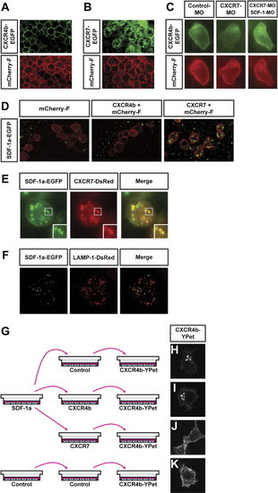
CXCR7 Is an SDF-1a Receptor that Promotes the Internalization of the Chemokine (A and B) Subcellular localization of CXCR4b and CXCR7 (green) in somatic cells of the embryo. CXCR4b (green) is predominantly found on the membrane of cells (red label of farnesylated mCherry) (A), while CXCR7 (green) is found on the plasma membrane and intracellularly (B). (C) CXCR7 knockdown increases extracellular SDF-1a levels as judged by internalization of CXCR4b in PGCs. In control embryos (left panel), CXCR4b (green) localizes to the plasma membrane of PGCs (red). CXCR7 knockdown leads to a reduction of CXCR4b on the membrane (middle panel). Membrane localization of CXCR4b in CXCR7 knockdown embryos is restored by SDF-1 knockdown (right panel). (D) SDF-1a is internalized by CXCR7-expressing cells. Somatic cells (red membrane) expressing CXCR7, CXCR4b, or a control protein were transplanted into host embryos that globally expressed SDF-1a-EGFP. Confocal images were taken 1 hr after transplantation. Transplanted cells (red) expressing either control protein or CXCR4b (left and middle panel, respectively) do not show uptake of SDF-1a (green). In contrast, cells expressing CXCR7 showed intracellular accumulations of SDF-1a protein (right panel). (E) SDF-1a and CXCR7 colocalize in vesicular structures. Images were taken 1 hr after transplantation of cells expressing CXCR7-DsRedMonomer into SDF-1a-EGFP-expressing hosts. The inset shows a magnification of the dotted box. (F) SDF-1a accumulates in lysosomes upon CXCR7-mediated internalization. Deconvoluted images were taken 1 hr after transplantation of cells expressing untagged CXCR7 and the lysosomal marker LAMP-1 fused to DsRedMonomer into SDF-1a-EGFP-expressing host embryos. (G–K) CXCR7-expressing cells reduce extracellular SDF-1a levels. In (G) is a graphic illustration of the experiments designed to examine the depletion of SDF-1a from conditioned medium by CXCR7-expressing cells. The conditioned medium was incubated with cells transfected with the different DNA constructs and subsequently transferred to reporter cultures expressing CXCR4b-EGFP. The extent of CXCR4b-EGFP internalization was then determined. In (H), strong CXCR4b internalization is observed in cells exposed to medium treated with control cells. In (I), medium depleted by CXCR4b-expressing cells induced CXCR4b internalization in 87.5% of all reporter cells, compared to control. CXCR4b ), strong CXCR4b internalization is observed in cells exposed to medium treated with control cells. In (I), medium deinternalization was only observed in 56.3% of cells exposed to medium depleted by CXCR7-expressing cells (J). |
Reprinted from Cell, 132(3), Boldajipour, B., Mahabaleshwar, H., Kardash, E., Reichman-Fried, M., Blaser, H., Minina, S., Wilson, D., Xu, Q., and Raz, E., Control of chemokine-guided cell migration by ligand sequestration, 463-473, Copyright (2008) with permission from Elsevier. Full text @ Cell