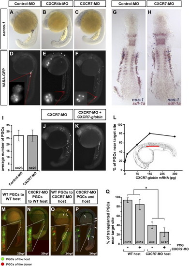
CXCR7 Is Essential for Normal PGC Migration and Is Required in the Somatic Environment of the Embryo (A–C) Reduction of CXCR7 activity leads to aberrant PGC migration as demonstrated by in situ hybridization using a germ cell-specific nanos1 probe. PGCs in control embryos cluster at the region where the gonad develops after 20 hr of development (A). Similar to reduction of CXCR4b activity (B), knockdown of CXCR7 results in a pronounced germ cell migration phenotype (C). (D–F) CXCR7 knockdown does not affect PGC specification. Images of 22 hpf embryos injected with Vasa-GFP-nos1-3′ UTR mRNA are shown. Germ cell-specific mRNA protection and proper localization of the Vasa-GFP fusion protein to germinal granules is observed in embryos injected with control morpholino (D), CXCR4b morpholino (E), and CXCR7 morpholino (F). (G and H) General embryonic patterning and expression of sdf-1a are not affected by CXCR7 knockdown. Two-color in situ hybridization using nanos1 (blue) and sdf-1a probes (red) of embryos injected with control (G) or CXCR7 (H) morpholino is shown. (I) CXCR7 knockdown does not affect PGC number as counted at 12 hpf. n signifies the number of embryos examined. Error bars represent standard error of the mean (SEM). (J–L) The effect of CXCR7 morpholino on PGC migration is reversed by CXCR7 expression. The severe migration phenotype induced by the CXCR7 antisense oligonucleotide (J) is reversed by global expression of RNA encoding CXCR7 (K). A graph demonstrating the dose-dependent rescue of the CXCR7 morpholino-induced phenotype by global expression of CXCR7 (L). For all injections the total amount of injected mRNA was identical (300 pg) by addition of control mRNA (mCherry-F-globin mRNA). The red bar indicates the correct target for the migrating PGCs. (M–Q) PGC migration depends on the activity of CXCR7 in somatic tissues. PGCs expressing DsRedExpress (red) were transplanted into embryos with PGCs expressing EGFP-F (green). Wild-type and CXCR7 knocked-down PGCs arrived at the region of the gonad in wild-type hosts (M and N). A large proportion of wild-type and CXCR7-depleted PGCs does not arrive at the correct target in CXCR7-depleted host embryos (O and P, arrowheads). In (Q), the percent of transplanted PGCs reaching their target after the first day of development isrge proportion of wild-type and CXCR7-depleted PGCs does not arrive at the correct target in CXCR7-depleted host embryos (O a shown. PGCs in CXCR7-depleted hosts embryos show a significant reduction of migration fidelity as compared to PGCs in wild-type hosts (p < 0.001, t test, marked with an asterisk). PGCs deficient for CXCR7 do not show a significant difference in arriving at the target as compared to control PG
|
