
Fig. 3
- ID
- ZDB-FIG-080325-60
- Publication
- Wilkins et al., 2008 - Mtx2 directs zebrafish morphogenetic movements during epiboly by regulating microfilament formation
- Other Figures
- All Figure Page
- Back to All Figure Page
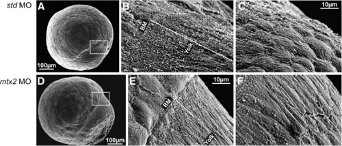
Scanning electron microscopy of the E-YSL. (A-F) Scanning electron microscopy of std MO and mtx2 MO injected embryos at 70-80% epiboly. The region between the blastoderm and the yolk (dashed box in panels A and D) is the site of the external yolk syncytial layer (E-YSL). 2000x magnification (B, C, E and F) reveals that std MO embryos (B, C) have extensive membrane folding in a region ~50-60 µm long (dashed line in panel B) that obscures the blastoderm margin/E-YSL border (″BM″ in panel B) and also the E-YSL/yolk border ("Yolk" in panel B). The membrane folding is most extensive closest to the blastoderm margin and decreases vegetally (C). In contrast, mtx2 MO embryos (E, F) have greatly reduced membrane folding, such that the margins of the E-YSL (″BM″ and ″yolk″ in panel E) can both be seen. Yolk membrane folding is also reduced (F). |
| Fish: | |
|---|---|
| Knockdown Reagent: | |
| Observed In: | |
| Stage Range: | Shield to 75%-epiboly |
Reprinted from Developmental Biology, 314(1), Wilkins, S.J., Yoong, S., Verkade, H., Mizoguchi, T., Plowman, S.J., Hancock, J.F., Kikuchi, Y., Heath, J.K., and Perkins, A.C., Mtx2 directs zebrafish morphogenetic movements during epiboly by regulating microfilament formation, 12-22, Copyright (2008) with permission from Elsevier. Full text @ Dev. Biol.