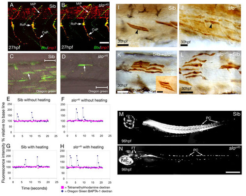
The phenotype of sloθ45 mutants is intrinsic to the muscle fibres. (A,B) Confocal projections of wild-type (A) and slou45 (B) embryos stained with alpha-bungarotoxin (Btx, green) to mark motor endplates and with the ZNP1 antibody to label primary motoneuron axons (znp1, red). The trajectories of the three primary motoneuron axons (MiP, medial; RoP, rostral; CaP, caudal) in the slou45 mutant (B) are similar to its sibling (A) with multiple bungarotoxin-positive puncta (neuromuscular junctions) associated with the axons in both cases. The only defect we detected was a slight reduction in the branching of some CaP axons in the ventral myotome of slou45 mutants. Bungarotoxin-positive spots are also evident in the furrow between the myotomes as noted previously (Behra et al., 2002; Lefebvre et al., 2007). (C-H) Calcium fluxes in the muscle of slou45 mutants are normal. (C,D) Muscle fibres (arrows) labelled with the calcium-sensitive dye Oregon-Green 488 BAPTA-1 dextran co-labelled with tetramethylrhodamine-dextran (to detect calcium-insensitive fluorescent changes) in myotomes of a slou45 mutant (D) and a sibling (C). Recordings of fluorescence in fibres from a mutant (F,H) and a sibling (E,G) with and without an external stimulus of heating show no differences except that transients are slightly larger in the mutant because movement in wild types took the dye-containing cells out of focus. To control for this movement, the ratio of peaks in calcium and rhodamine fluorescence was calculated (see Table S1). (I-L) Muscle fibres of slou45 mutants have abnormal morphologies. (I,J) Mosaic labelling of muscle cells by injection of biotinylated dextran and ABC Kit (Vector labs) and DAB in a sibling (I) and a slou45 mutant (J). slou45 muscle fibres have an inconsistent, uneven and wavy morphology lacking crisp attachments at the myotome boundaries, in contrast to the regimented, straight and crisply attached appearance of muscle fibres in siblings. (K,L) The slou45 mutation has a cell-autonomous effect. Dextran-labelled cells were transplanted from/to wild-type and slou45 embryos at 30% epiboly, fixed and stained at 24-36 hpf. Most wild-type sibling muscle cells show near-normal morphology in a slou45 mutants (K), although isolated wild-type cells tended to have a slightly defective morphology - when the cells grouped together (inset), their morphology is more normal. slou45 mutant cells in a wild-type sibling show abnormal morphology (L) similar to non-transplanted mosaically labelled mutant cells (compare with J). This suggests that the slo phenotype is primarily due to a cell-autonomous defect with only minor, likely secondary, non-autonomous defects. (M,N) Lateral views of trunk muscles of a slou45 mutant (N) and a sibling (M) visualised with polarised light. Muscle fibres in the wild type are highly birefringent, whereas in the slou45 mutant, the muscle completely lacks birefringence. Birefringence is still seen in the pigment cells (PC), eye (E) and in fibre-tracts of the brain (FT) and appears brighter in mutants due to longer photographic exposure. Scale bars: 20 μm in A,B,I-L; 50 μm in C,D; 200 μm in M,N.
|
