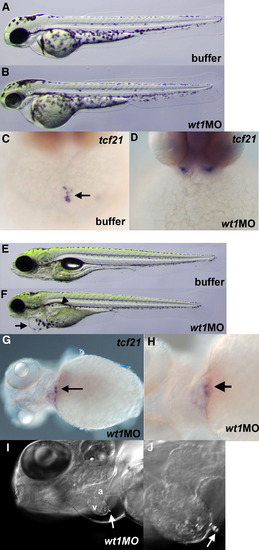
wt1 is required for development of the PEO. Live 48 hpf embryos injected with buffer (A) as a control or with wt1MO (B). A slight pericardial edema is observed in the wt1MO embryos. tcf21 staining at 48 hpf of (C) buffer-injected or (D) wt1MO-injected embryos. The PEO is clearly visible in the control embryos (arrow) but is completely lacking in wt1MO embryos (127/127, 100%). (E) Control and (F) wt1MO-injected embryos shown at 4 dpf. wt1MO embryos display pericardial edema (arrow in panel F) as well as edema in the pronephros (arrowhead). (G) At 4 dpf, a fraction of the embryos (22/68, 32%) have tcf21-positive cells adjacent to the heart (arrow). (H) Same embryo as panel G but shown at higher magnification with the wt1-positive cells (arrow) nestled between the heart and yolk as observed in 48 hpf wild-type embryos. (I) Live view of a 4 dpf wt1MO embryo using DIC optics. The ventricle (v) and atrium (a) are marked and an arrow points to a PEO-like cluster on the surface of the ventricular myocardium. (J) Higher magnification view of the embryo in panel I.
|
