
Fig. 1
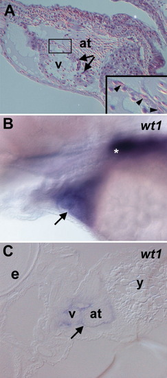
The epicardium in the larval zebrafish heart expresses wt1. (A) Hematoxylin and eosin-stained section of a 4 dpf zebrafish embryo detailing the structure of the embryonic heart. The atrium (at) has a thinner wall than the ventricle (v) and valves are present between the two chambers (arrows). Myocardial nuclei are round while epicardial nuclei (arrowheads in the inset) appear elongated. (B) Whole-mount in situ hybridization of a 4 dpf embryo with the wt1 probe. The pronephric expression is marked with an asterisk (*) and cardiac staining (arrow) is clearly visible. (C) Frontal section of a 4 dpf wt1-stained embryo confirming that the wt1 expression is localized to the outer cardiac layer (arrow). Anterior is to the left in all panels. e: eye, y: yolk. |
| Gene: | |
|---|---|
| Fish: | |
| Anatomical Term: | |
| Stage: | Day 4 |
Reprinted from Developmental Biology, 315(1), Serluca, F.C., Development of the proepicardial organ in the zebrafish, 18-27, Copyright (2008) with permission from Elsevier. Full text @ Dev. Biol.