
Fig. 5
- ID
- ZDB-FIG-080324-13
- Publication
- Sullivan-Brown et al., 2008 - Zebrafish mutations affecting cilia motility share similar cystic phenotypes and suggest a mechanism of cyst formation that differs from pkd2 morphants
- Other Figures
- All Figure Page
- Back to All Figure Page
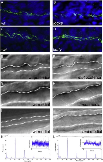
Early pronephric cyst phenotypes. (A–D) Acetylated tubulin staining in the pronephric tubules at 27 hpf. Cilia were detected using an anti-acetylated tubulin antibody (green) and counterstained with the nuclear marker Hoechst (blue). Cilia appeared grossly normal in swt and kurly, but are shorter in locke when compared to wild-type siblings. (E–J) DIC microscopy images of the pronephric tubules in wild-type siblings and kurly mutants. Lumen sizes in posterior tubules from 26–30 hpf are similar in both wild-type siblings (E) and mutant embryos (F). Lumens in the medial tubules are larger in both wild-type siblings (G) and mutant embryos (H) when compared in the posterior regions (E, F). By 2 dpf, there is a clear dilation in the medial tubules of mutant embryos (J) as compared to wild-type siblings (I). (K, L) Infra-red scattering measurements demonstrate that the frequency of cilia movement in kurly mutants (L) is similar to the frequency observed in wild-type sibling embryos (K) (∼ 42 Hz). Both spectra show several peaks. The measurements demonstrate that the frequency of cilia movement in kurly mutants (L) is similar to the frequency observed in wild-first peak in each spectrum corresponds to the fundamental frequency of cilia movement. The subsequent peaks corresponds to the harmonics of the fundamental frequency (∼ n * 42 Hz with n = 1, 2 and 3). Panels E–J were taken with a 60x objective water immersion lens using two different cameras. Panels E–H were taken with an iXon camera (Andor); pixel size 16 * 16 μm. Panels I and J were taken with a Luca camera (Andor); pixel size of 10 * 10 μm. Because of differences in pixel size, luminal size in panels E–H cannot be directly compared to panels I and J. |
| Fish: | |
|---|---|
| Observed In: | |
| Stage Range: | Prim-5 to Long-pec |
Reprinted from Developmental Biology, 314(2), Sullivan-Brown, J., Schottenfeld, J., Okabe, N., Hostetter, C.L., Serluca, F.C., Thiberge, S.Y., and Burdine, R.D., Zebrafish mutations affecting cilia motility share similar cystic phenotypes and suggest a mechanism of cyst formation that differs from pkd2 morphants, 261-275, Copyright (2008) with permission from Elsevier. Full text @ Dev. Biol.