
Fig. S5
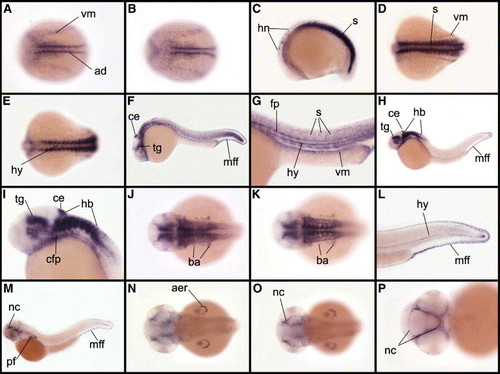
Expression pattern of follistatin-like 1 during embryonic development. (A, B) Expression of fstl1 starts at the beginning of somitogenesis in adaxial cells (ad), anterior somites and ventral mesenchyme (vm). (C–E) At the 15-somite stage, expression of fstl1 is observed in somites (s) and hindbrain neurons (hn), in ventral mesoderm (vm) as well as in hypochord (hy). At 24 hpf (F–G), expression is observed in tegmentum (tg), cerebellum (ce), hindbrain, floor plate (fp) and hypochord in median fin fold (mff). A weak expression is also observed in somites and in ventral mesenchyme (vm). At 36 hpf (H–L), fstl1 is expressed in tegmentum, cephalic floor plate (cfp) and dorsal part of hypothalamus, in cerebellum, in hindbrain, in a subpopulation of cell of branchial arches (ba), hypochord and median fin fold. At 48 hpf (M–P), fstl1 is expressed in neurocranium (nc), central part of pectoral fins (pf), apical ectodermal ridge of pectoral fins (aer), caudal hypochord and median fin folds. Embryos in dorsal views, anterior to the left in A, B, D, E, J, K and N–P and in lateral views, anterior to the left in C, F–I, L and M. |
| Gene: | |
|---|---|
| Fish: | |
| Anatomical Terms: | |
| Stage Range: | 1-4 somites to Long-pec |
Reprinted from Developmental Biology, 298(2), Dal-Pra, S., Fürthauer, M., Van-Celst, J., Thisse, B., and Thisse, C., Noggin1 and Follistatin-like2 function redundantly to Chordin to antagonize BMP activity, 514-526, Copyright (2006) with permission from Elsevier. Full text @ Dev. Biol.