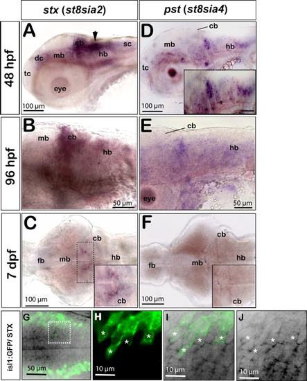
stx and pst expression in the differentiating zebrafish central nervous system. Panels A,B,D,E show lateral and C,F dorsal views, anterior is to the left. A: At 48 hours postfertilization (hpf) stx expression is weakly present in the ventral telencephalon, in the diencephalon, midbrain, in the floorplate, and in the spinal cord. Strong expression is found in dorsal regions of the caudal hindbrain along the fourth ventricle (black arrow) and in the differentiating cerebellum. B: stx expression remains weakly detectable in the differentiating cerebellum and in ventral regions of the mid- and hindbrain at 96 hpf. C: At 7 days postfertilization (dpf), only the dorsomedial domain of the hindbrain retains low levels of stx (dashed box and inset showing high magnification of the dorsomedial domain in the anterior hindbrain with remaining stx expression). D: At 48 hpf, pst is expressed in the ventral domains of the fore- and midbrain. A striped expression pattern can be observed in the hindbrain, being confined to the central domains of individual rhombomeres (inset, asterisks). E: Only some patches in the hindbrain retain low pst expression levels at 96 hpf. F: pst expression is no longer detectable in the brain at 7 dpf (compare inset in F with inset in C). G-J: Expression of stx in a subset of cranial motoneurons as revealed by colabeling of stx and green fluorescent protein (GFP) in embryos of the islet1:GFP transgenic line. G: Maximum intensity projection of a 30-μm stack of optical sections showing stx expression in medial domains of the hindbrain (black) and more laterally positioned GFP-expressing cranial motoneurons (green). H-J: A subset of cranial motoneurons (H, white asterisks) expresses stx (J, white asterisks), which can be identified in 1-μm single optical sections at high magnification. I: Overlay of GFP and stx expression. cb, cerebellum; dc, diencephalon; fb, forebrain; hb, hindbrain; mb, midbrain; sc, spinal cord; tc, telencephalon.
|
