
Fig. 4
- ID
- ZDB-FIG-071219-8
- Publication
- Bertrand et al., 2007 - Definitive hematopoiesis initiates through a committed erythromyeloid progenitor in the zebrafish embryo
- Other Figures
- All Figure Page
- Back to All Figure Page
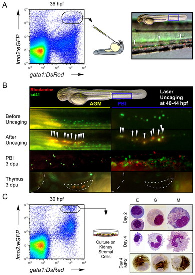
Functional studies demonstrate that gata1+ lmo2+ CD41+ cells are committed erythromyeloid progenitors. (A) Dissociated gata1:DsRed+ lmo2:eGFP+ cells were purified from 36 hpf embryos by flow cytometry and transplanted into wild-type embryonic recipients. Transplanted cells were observed to home back to the PBI in host animals (right panel, 200x magnification). Arrowheads denote gata1:DsRed+ lmo2:eGFP+ cells. (B) In vivo fate-mapping studies were performed by laser activation of caged rhodamine in CD41+cells in the 44 hpf AGM or 40 hpf PBI. Presumptive AGM HSCs were targeted as positive controls for thymus colonization (lower panels; outlined crescent-shaped structure). Boxed yellow and blue regions in upper panel denote close-up areas shown below in left panels for the AGM and right panels for the PBI, respectively. All animals are shown in lateral view, with head oriented to the left and dorsal side up. Dotted line at the left edge of lower boxes denotes outline of the eye for orientation. White arrowheads indicate uncaged, GFP+ cells. (C) Short-term culture of lmo2+ gata1+ cells atop kidney stromal cells demonstrates erythroid (E), granulocytic (G) and monocytic/macrophage (M) differentiation potentials. Cultured cells were stained for myeloperoxidase (MPX) activity after 4 days (bottom panel). |