
Fig. 5
- ID
- ZDB-FIG-071219-17
- Publication
- Bertrand et al., 2007 - Definitive hematopoiesis initiates through a committed erythromyeloid progenitor in the zebrafish embryo
- Other Figures
- All Figure Page
- Back to All Figure Page
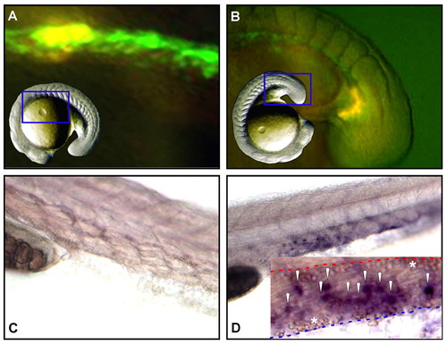
Erythromyeloid progenitors arise autonomously within the PBI. (A-D) Cells expressing GFP under control of the lmo2 promoter were lineage traced by uncaging a combination of caged rhodamine and FITC. GFP+ cells were targeted, either in the medial (bounded by somites 1-10; panel A) or most posterior (panel B) regions of the lmo2 expression domain between 13- and 15-somite stages. (C,D) Analysis of targeted progeny at 30 hpf showed no medially derived cells in the PBI (C), whereas posterior-derived daughter cells were observed throughout the venous plexus of the PBI (D). Inset in (D) shows a close-up of the PBI in a second animal, with marked progeny found within the vascular plexus between the aorta (dashed red line) and caudal vein (dashed blue line). Primitive erythrocytes within each vessel are indicated with asterisks and the daughters of posterior lmo2+ cells are indicated by white arrowheads. Blue framed areas in inset images of whole animals indicate the regions shown in close-up in A and B. |