
Fig. 6
- ID
- ZDB-FIG-071127-16
- Publication
- Abe et al., 2007 - Function of FGF signaling in the developmental process of the median fin fold in zebrafish
- Other Figures
- All Figure Page
- Back to All Figure Page
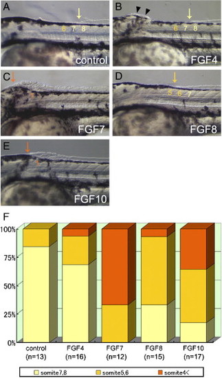
Ectopic median fin fold-like structure formation was induced by FGF application to the dorsal midline. (A–E) Lateral view of the anterior trunk region in 42 hpf or 24 h post implantation embryos. FGF-soaked beads were implanted into the dorsal midline at around the second and third somite level of 18 hpf embryos. Arrows indicate the anterior edge of the median fin fold and ectopic median fin fold-like structure. (A) Control embryos usually had the anterior end of the median fin fold at the 8th somite level. (B–E) Ectopic median fin fold-like structure could be seen rostrally to the intrinsic median fin fold after implantation of FGF beads (B; FGF4, C; FGF7, D; FGF8, E; FGF10). Arrowheads in panel B indicate ectopic hypertrophy that was disconnected from the intrinsic fin fold. (F) The ratio of the position of the anterior end of the median fin fold-like structures in the bead-implanted embryos. The colors of bars in the graph indicate the position of the anterior end in the median fin fold and the median fin fold-like structure: yellow is somite 7–8 level, orange is somite 5–6 level, and maroon is somite 4< region. The colors of bars correspond to the colors of arrows in panels A–E. |
Reprinted from Developmental Biology, 304(1), Abe, G., Ide, H., and Tamura, K., Function of FGF signaling in the developmental process of the median fin fold in zebrafish, 355-366, Copyright (2007) with permission from Elsevier. Full text @ Dev. Biol.